Nuclear Medicine Technologist
March 29, 2024
Updated April 13, 2025
18 minute read
Exploring a Career as a Nuclear Medicine Technologist
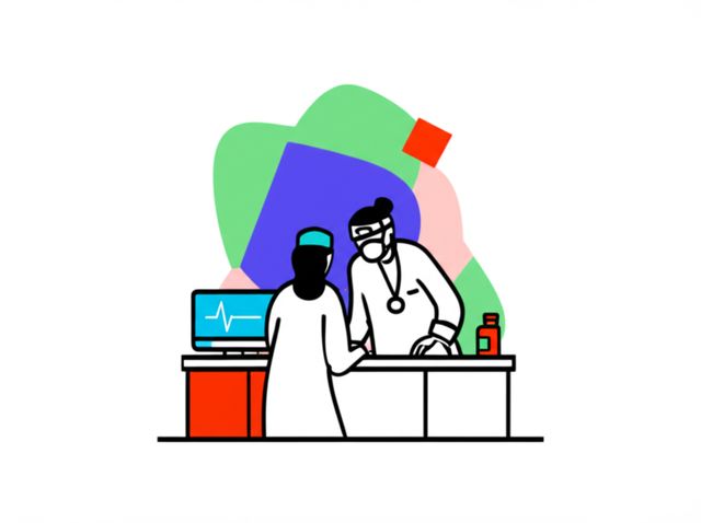
A Nuclear Medicine Technologist is a specialized healthcare professional who works directly with patients and physicians, using radioactive materials, known as radiopharmaceuticals, to diagnose and treat diseases. They operate sophisticated imaging equipment that traces the path of these substances within a patient's body, creating images that reveal how organs and tissues are functioning. This field combines patient care, advanced technology, and a deep understanding of physics and biology.
16cp7x|
Find a path to becoming a Nuclear Medicine Technologist. Learn more at:
OpenCourser.com/career/16cp7x/nuclear
Reading list
We haven't picked any books for this reading list yet.
Provides a comprehensive overview of the use of PET and SPECT imaging in oncology. It covers a wide range of topics, including the principles of these imaging techniques, their clinical applications, and their role in the diagnosis and management of cancer.
A comprehensive reference on the principles, artifacts, and advanced techniques of x-ray imaging and computed tomography.
Provides a comprehensive overview of the theory and practice of PET.
A comprehensive textbook that covers the fundamental principles, technical aspects, and clinical applications of x-ray imaging.
Covers the use of PET and SPECT in the diagnosis and management of cancer. The book includes chapters on the use of PET and SPECT in the diagnosis of lung cancer, breast cancer, colorectal cancer, and prostate cancer.
Provides a comprehensive overview of the physics of medical imaging, including X-ray imaging, computed tomography, and magnetic resonance imaging. It is suitable for students, researchers, and practitioners in the field of medical imaging.
A practical guide to X-ray imaging in medical applications, this book provides detailed coverage of the underlying physics, instrumentation, and clinical techniques.
A broad overview of x-ray imaging techniques and their applications in various fields, including medical imaging, industrial inspection, and security.
Provides a broad overview of the principles of medical imaging, covering all major imaging modalities used in clinical practice. It is written in a clear and concise style, making it suitable for students and practitioners alike.
Provides a comprehensive overview of the physics underlying medical imaging techniques, including PET. It is written in a clear and concise style and is suitable for students and practitioners with a basic understanding of physics.
Provides a comprehensive overview of the use of PET and SPECT in molecular imaging, covering principles, techniques, and applications in various biological processes and diseases.
This textbook comprehensive guide to the principles and practice of surgery, including a detailed section on surgical radiology.
A specialized book that focuses on the medical applications of x-ray imaging, including radiography, fluoroscopy, and computed tomography.
Explores the use of PET and SPECT in oncology, covering tumor biology, radiopharmaceutical development, clinical applications, and future directions.
An exploration of the historical development of X-ray imaging, this book offers insights into the scientific and technological breakthroughs that have shaped the field.
A specialized book that focuses on the principles and applications of x-ray optics, which is essential for understanding the design and performance of x-ray imaging systems.
A specialized book that focuses on the use of x-ray imaging in biomedical research, including techniques such as micro-CT and nano-CT.
A specialized book that focuses on the security applications of x-ray imaging, including cargo scanning, baggage inspection, and border security.
Provides a comprehensive overview of the use of PET-CT imaging in clinical oncology, covering the basic principles, clinical applications, and future directions. It is written by two leading experts in the field and is essential reading for anyone interested in this topic.
Provides a comprehensive overview of nuclear medicine therapy, including the use of PET for treatment planning and monitoring.
A classic textbook that covers the principles and applications of x-ray diffraction crystallography, which related technique to x-ray imaging.
Is written for practitioners who are interested in using PET-CT in clinical practice. It covers the basic principles of PET-CT, as well as the clinical applications of PET-CT in a variety of diseases. It is written by a team of experienced clinicians and researchers.
Provides a detailed overview of the imaging anatomy and pathology of the head and neck, including the brain, skull, and facial structures.
Provides a comprehensive overview of the use of PET imaging in cancer, covering the basic principles, clinical applications, and future directions. It is written by a team of experienced clinicians and researchers.
For more information about how these books relate to this course, visit:
OpenCourser.com/career/16cp7x/nuclear