March 29, 2024
Updated May 12, 2025
22 minute read
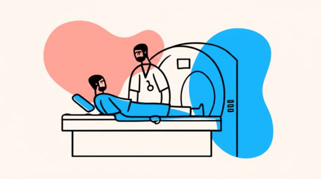
A Radiologic Technologist, often called an R.T. or radiographer, is a healthcare professional who performs diagnostic imaging examinations on patients. They use various types of sophisticated medical equipment to create images of the inside of the human body, which are then interpreted by physicians, primarily Radiologists, to diagnose and monitor diseases or injuries. This crucial role places Radiologic Technologists at the forefront of patient care, directly impacting diagnostic accuracy and treatment planning.
Working as a Radiologic Technologist can be engaging and exciting for several reasons. Firstly, it's a field that blends direct patient interaction with advanced technology. You'll be responsible for explaining procedures to patients, ensuring their comfort, and precisely positioning them for imaging, all while operating complex machinery. Secondly, the field offers diverse specialization opportunities, allowing you to focus on areas like mammography, computed tomography (CT), or magnetic resonance imaging (MRI), each with unique challenges and rewards. Finally, the work of a Radiologic Technologist is fundamental to modern medicine; the images they produce are often the key to unlocking a diagnosis and guiding effective treatment, making it a deeply impactful career.
What is a Radiologic Technologist?
Radiologic Technologists are integral members of the healthcare team who specialize in performing medical imaging procedures. Their primary responsibility involves using specialized equipment to capture images of patients' internal body structures. These images are critical for physicians to diagnose and treat a wide array of medical conditions, from bone fractures to complex diseases. It's important to distinguish that while "Radiologic Technician" is an older term, "Radiologic Technologist" is the more current and accurate title for this profession.
lukikj|
Find a path to becoming a Radiologic Technologist. Learn more at:
OpenCourser.com/career/lukikj/radiologic
Featured in The Course Notes
This career is mentioned in our blog,
The Course Notes. Read
two articles that feature
Radiologic Technologist:
To read more articles from OpenCourser, visit:
OpenCourser.com/notes
Reading list
We haven't picked any books for this reading list yet.
This landmark report from the Institute of Medicine was the first to quantify the problem of medical errors in the United States. It has had a profound impact on the way that we think about and approach patient safety.
Classic text on the art of diagnosis. It must-read for students and practitioners alike.
Provides a comprehensive overview of internal medicine. It is an excellent resource for medical students, residents, and practicing physicians.
Provides a comprehensive overview of the rational clinical examination. It valuable resource for students and practitioners alike.
Provides a comprehensive guide to the clinical examination. It valuable resource for students and practitioners alike.
Provides a comprehensive overview of artificial intelligence in clinical diagnosis. It valuable resource for students and practitioners alike.
Provides a comprehensive overview of clinical decision making. It valuable resource for students and practitioners alike.
This comprehensive textbook covers all aspects of radiation therapy and oncology. It is written by Stephen Chmura, who leading expert in the field. The book covers the basic principles and techniques of radiation therapy and oncology, as well as the clinical applications of these technologies.
Provides a comprehensive overview of current diagnosis and treatment of common surgical conditions.
This comprehensive textbook covers all aspects of sonography. It is written by Frederick Kremkau, who leading expert in the field. The book covers the basic principles and techniques of sonography, as well as the clinical applications of this technology.
This report from the National Patient Safety Foundation outlines a vision for the future of patient safety. It identifies six key areas for improvement, including medication safety, infection prevention, and the use of technology.
This concise guide provides a quick reference to the key concepts and techniques in diagnostic imaging. It is ideal for clinicians who need a quick and easy way to refresh their knowledge or for students who are just starting to learn about this topic.
Provides a practical guide to diagnosing common medical conditions based on their symptoms. It useful resource for students and practitioners alike.
Provides a practical guide to biostatistics in clinical medicine. It useful resource for students and practitioners alike.
By Atul Gawande, a surgeon and writer, explores the power of checklists to reduce errors in medicine and other fields. It fascinating and inspiring read for anyone who is interested in improving safety and efficiency.
This report from the Institute of Medicine outlines a plan for eliminating preventable deaths from cancer. It focuses on the importance of early detection, prevention, and access to care.
Explores the cultural factors that contribute to medical errors. Dekker argues that safety is not simply a matter of following rules and procedures, but also of creating a culture in which people feel comfortable speaking up about errors and learning from them.
Provides a detailed overview of the musculoskeletal system as seen through various imaging modalities. It is written by a team of experts in radiology and musculoskeletal anatomy and is well-illustrated with high-quality images.
Provides a comprehensive overview of computed tomography (CT), covering the principles of the technology, the different types of CT scanners, and the clinical applications of CT. It is written by a team of experts in the field and is well-illustrated with high-quality images.
This introductory textbook good starting point for students who are new to diagnostic imaging. It covers the basic principles and techniques of the field and includes case studies to help readers apply their knowledge to real-world situations.
Argues that the healthcare system is ripe for disruption by new technologies and business models. It offers a number of specific recommendations for how to improve the quality and affordability of care.
Provides a concise overview of magnetic resonance imaging (MRI), covering the principles of the technology, the different types of MRI scanners, and the clinical applications of MRI. It is written by an expert in the field and is well-illustrated with high-quality images.
Provides a basic overview of diagnostic imaging for non-radiologists. It covers the different types of imaging modalities and their clinical applications. It is written by a team of experts in the field and is well-illustrated with high-quality images.
Provides a comprehensive overview of diagnostic imaging of the head and neck, covering the different types of imaging modalities and their clinical applications in the diagnosis and management of head and neck diseases. It is written by a team of experts in the field and is well-illustrated with high-quality images.
For more information about how these books relate to this course, visit:
OpenCourser.com/career/lukikj/radiologic

