We may earn an affiliate commission when you visit our partners.

Medical Imaging Analyst

Save
April 2, 2024 Updated April 7, 2025 15 minute read

Medical Imaging Analyst: Shaping the Future of Healthcare Through Data

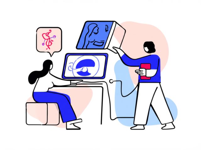
Medical Imaging Analysts operate at the exciting intersection of healthcare, technology, and data science. They are specialized professionals who focus on processing, analyzing, and interpreting complex medical images generated by various technologies like MRI, CT, and PET scans. Their work goes beyond simply viewing images; it involves applying computational techniques, often including artificial intelligence (AI), to extract quantitative information, identify patterns, assist in diagnoses, plan treatments, and contribute to medical research. Think of them as translators, turning visual data into actionable insights for doctors and scientists.

Working in this field means playing a crucial role in patient care and medical discovery. You might be involved in developing algorithms that help detect diseases earlier, creating detailed 3D models for surgical planning, or managing vast datasets for clinical trials. It's a dynamic career that demands both technical expertise and a strong understanding of medical contexts, offering the chance to directly impact health outcomes and push the boundaries of medical technology.

Introduction to Medical Imaging Analysis

What is Medical Imaging Analysis?

Medical Imaging Analysis is a specialized field focused on extracting meaningful information from medical images using computational tools and techniques. Analysts in this role apply sophisticated software and algorithms to process images from modalities like X-ray, Computed Tomography (CT), Magnetic Resonance Imaging (MRI), Positron Emission Tomography (PET), and Ultrasound. Their goal is to enhance image quality, identify features, quantify measurements, and ultimately support clinical decisions or research objectives.

The scope extends beyond routine image viewing performed by radiologists. Analysts often engage in tasks like image segmentation (isolating specific organs or tissues), registration (aligning different images), and feature extraction (measuring characteristics like size, shape, or texture). This quantitative data provides deeper insights than visual inspection alone.

Essentially, medical imaging analysis bridges the gap between raw image data and clinical or research application. It requires a blend of knowledge in anatomy, physiology, physics (of imaging), computer science, and data analysis to transform pixels into knowledge.

Key Applications in Healthcare

The applications of medical imaging analysis are vast and vital across healthcare. In diagnostics, analysts help refine image data, making subtle abnormalities easier for radiologists to spot. They might develop or use AI tools that flag suspicious areas in scans, potentially leading to earlier cancer detection or more accurate assessments of neurological disorders.

For treatment planning, especially in surgery and radiation oncology, analysts create detailed 3D reconstructions of patient anatomy from scans. These models allow surgeons to precisely plan procedures or radiation therapists to target tumors while minimizing damage to surrounding healthy tissue. This personalized approach significantly improves treatment efficacy and safety.

In research, medical imaging analysis is indispensable. Analysts process and quantify imaging data from clinical trials to objectively measure treatment effects or track disease progression. Their work helps validate new therapies and deepen our understanding of various diseases, contributing to the evidence base that drives medical advancements.

Related Fields and Technologies

Medical Imaging Analysis doesn't exist in isolation; it's closely intertwined with several other fields. Radiology is the most obvious connection, as analysts often work alongside radiologists, supporting their diagnostic interpretations with quantitative data and advanced visualizations. However, the analyst focuses more on the computational processing and data extraction, whereas the radiologist provides the final clinical diagnosis.

Bioinformatics is another related area, particularly as studies increasingly integrate imaging data with genomic, proteomic, or other biological data. Analysts may collaborate with bioinformaticians to correlate imaging features with genetic markers, contributing to personalized medicine.

Perhaps the most significant influence currently is Artificial Intelligence (AI), particularly deep learning. AI is revolutionizing image analysis by enabling automated detection, segmentation, and classification tasks with increasing accuracy. Medical Imaging Analysts are often involved in developing, validating, implementing, and monitoring these AI tools within clinical or research workflows.

These connections highlight the interdisciplinary nature of the role, requiring a broad understanding of both medical and technological domains.

Key Responsibilities of a Medical Imaging Analyst

Core Tasks and Workflows

A primary responsibility of a Medical Imaging Analyst involves processing raw image data acquired from scanners. This includes tasks like noise reduction, contrast enhancement, and correcting for imaging artifacts to improve image quality for interpretation or further analysis. They apply various computational techniques, such as segmentation to delineate anatomical structures or lesions, and registration to align images taken at different times or from different modalities.

Analysts often manage complex analysis workflows, which might involve running specialized software or custom scripts. They ensure data integrity throughout the process, from initial acquisition to final reporting. Documenting methods and results meticulously is crucial for reproducibility and compliance.

Quantitative analysis is a key output. This could involve measuring tumor volumes, assessing blood flow, quantifying tissue characteristics, or extracting radiomic features (complex data patterns invisible to the eye). These objective measurements provide valuable information beyond qualitative visual assessment.

To get a hands-on feel for image processing tasks, exploring tools commonly used in the field can be beneficial. These courses offer introductions to core concepts and software.

Collaboration and Communication

Medical Imaging Analysts rarely work in isolation. Effective collaboration with radiologists and other clinicians is essential. Analysts provide technical expertise, helping clinicians understand the capabilities and limitations of different analysis techniques and interpreting the quantitative results in a clinical context.

They often act as a bridge between clinical teams and technical experts like software developers or AI engineers. Communicating clinical needs to technical teams and explaining technical complexities to clinicians requires strong interpersonal and translation skills. They might participate in multidisciplinary team meetings to discuss patient cases or research findings.

Documentation and reporting are also key communication tasks. Analysts must clearly present their methods, findings, and any limitations in reports or presentations tailored to different audiences, ranging from clinical colleagues to research collaborators or regulatory bodies.

Ensuring Quality and Managing Data

Maintaining high standards of quality assurance (QA) is paramount. Analysts perform checks on incoming image data to ensure it meets protocol requirements and is suitable for analysis. They also validate the performance of analysis tools and algorithms, ensuring they produce accurate and reliable results.

Data management is another critical responsibility. Medical imaging datasets can be enormous and complex. Analysts are often involved in organizing, archiving, and retrieving this data, frequently using Picture Archiving and Communication Systems (PACS) and adhering to standards like DICOM (Digital Imaging and Communications in Medicine).

Compliance with data privacy regulations, such as HIPAA in the US or GDPR in Europe, is non-negotiable. Analysts must ensure that patient data is handled securely and anonymized appropriately, especially when used for research or algorithm development.

Algorithm Development and Validation

In many roles, particularly in research or industry settings, Medical Imaging Analysts contribute to the development and validation of new analysis algorithms. This often involves leveraging AI and machine learning (ML), especially deep learning, to automate or improve tasks like disease detection, segmentation, or prediction.

Analysts might write code (commonly in Python or MATLAB), adapt existing algorithms, or utilize specialized software platforms. They design validation studies to rigorously test algorithm performance against ground truth data (e.g., expert annotations or clinical outcomes), assessing accuracy, robustness, and potential biases.

Staying abreast of the latest advancements in computer vision and AI is crucial for this aspect of the role. They need to understand the principles behind these algorithms to apply them effectively and critically evaluate their suitability for specific medical applications.

Developing skills in deep learning and its application to computer vision is increasingly important. These courses provide foundational knowledge and practical experience in this area.

For those serious about mastering the theoretical and practical aspects of AI/ML, these books are highly recommended resources.

Formal Education Pathways

Building the Foundation: Undergraduate Studies

A strong foundation typically begins with a bachelor's degree in a relevant scientific or technical field. Common majors include Biomedical Engineering, Computer Science, Physics, Mathematics, or Electrical Engineering. Coursework in biology, anatomy, and physiology is also highly beneficial to understand the clinical context.

Regardless of the specific major, core competencies should include strong analytical and problem-solving skills, proficiency in mathematics (calculus, linear algebra, statistics), and foundational programming knowledge. Exposure to image processing concepts or signal processing during undergraduate studies is a plus.

Some universities may offer specific tracks or minors related to medical imaging or bioinformatics, which can provide a more tailored preparation. Internships or research projects involving medical image analysis during undergraduate years can significantly strengthen an application for graduate studies or entry-level positions.

Advanced Studies: Graduate Programs

While some entry-level positions might be accessible with a bachelor's degree and relevant experience, many roles, especially in research and development, require a Master's or PhD degree. Graduate programs in Biomedical Engineering, Medical Physics, Computer Science (with a focus on AI/computer vision), or specialized Medical Imaging programs are common routes.

Master's programs often provide more specialized technical training and project experience, preparing graduates for industry or advanced technical roles. PhD programs are research-intensive, suitable for those aiming for careers in academia or leading research and development in industry. Research topics might include developing novel imaging techniques, creating advanced AI algorithms for specific clinical problems, or exploring new applications of imaging analysis.

Choosing a graduate program often involves considering faculty research areas, available resources (imaging equipment, computational facilities), and industry connections. It represents a significant investment, so careful research is warranted.

Understanding the fundamentals of various imaging modalities is essential. This course provides a broad overview suitable for those starting their journey.

These books offer in-depth coverage of the physics and principles underlying medical imaging technologies.

Certifications and Credentials

While formal degrees are the primary educational route, professional certifications can enhance credentials and demonstrate specialized expertise. The most relevant certification for this field is often the Certified Imaging Informatics Professional (CIIP), offered by the American Board of Imaging Informatics (ABII), which is a collaboration between the Society for Imaging Informatics in Medicine (SIIM) and the American Registry of Radiologic Technologists (ARRT).

record:16

The CIIP certification validates knowledge in areas crucial to imaging informatics, including PACS administration, image data management, IT systems, and workflow optimization. While not always mandatory, it can be advantageous for roles involving system administration or informatics management within healthcare institutions.

Other certifications related to specific software platforms, programming languages, or IT skills (like CompTIA certifications mentioned in some job postings) might also be beneficial depending on the specific role requirements. Continuous professional development and staying updated with technological advancements are generally expected, whether through formal certification or other learning avenues.

Technical Skills and Tools

Understanding Imaging Modalities

A fundamental requirement is understanding the principles behind various medical imaging modalities. This includes knowing how images are generated by technologies like Magnetic Resonance Imaging (MRI), Computed Tomography (CT), Positron Emission Tomography (PET), Single Photon Emission Computed Tomography (SPECT), Ultrasound, and X-ray.

This knowledge isn't just theoretical; it informs how analysts approach image processing and analysis. Understanding the physics helps identify potential artifacts, appreciate limitations in resolution or contrast, and choose appropriate analysis techniques for data from different sources. For example, processing MRI data requires different considerations than processing CT data.

Familiarity extends to understanding the typical clinical uses of each modality – which technique is best suited for imaging soft tissues versus bones, or for assessing function versus structure. This context is crucial for interpreting results correctly.

This course provides a foundational understanding of various medical imaging techniques.

These books delve deeper into the physics and technology behind medical imaging.

Essential Software and Platforms

Proficiency with specialized software is essential. Analysts regularly use Picture Archiving and Communication Systems (PACS) for accessing and managing images, and DICOM viewers for visualizing them. Familiarity with the DICOM standard itself is crucial for handling medical image data.

Beyond standard viewers, analysts use various image processing and analysis software platforms. These can range from open-source tools like ImageJ/Fiji and 3D Slicer to commercial packages specific to certain modalities or applications. Research environments might utilize platforms developed in-house.

Experience with data management tools and databases may also be required, especially in roles involving large-scale research projects or clinical data warehouses. Understanding workflow orchestration tools can also be beneficial for managing complex analysis pipelines.

This guided project helps learners get started with a popular open-source tool.

These books cover digital image processing principles often implemented in these software tools.

Programming and Computational Skills

Strong programming skills are increasingly vital, particularly in roles involving algorithm development, data analysis, or workflow automation. Python has become a dominant language in the field due to its extensive libraries for scientific computing, data analysis (NumPy, Pandas), image processing (Scikit-image, OpenCV), and machine learning (TensorFlow, PyTorch).

MATLAB remains widely used, especially in academic research and engineering contexts, offering powerful toolboxes for image processing and analysis. Depending on the specific environment and tasks, knowledge of other languages like C++ (for performance-critical applications) or R (for statistical analysis) can also be valuable.

Beyond specific languages, understanding fundamental computer science concepts, data structures, algorithms, and software development practices (like version control with Git) is important for developing robust and maintainable analysis tools.

These courses focus on applying deep learning using common programming frameworks.

These books provide practical guidance on using key programming libraries for machine learning and computer vision.

AI and Machine Learning in Imaging

Expertise in Artificial Intelligence (AI) and Machine Learning (ML), particularly deep learning, is becoming a core requirement for many Medical Imaging Analyst roles. This involves understanding concepts like convolutional neural networks (CNNs), recurrent neural networks (RNNs), and transformers, which are foundational to modern computer vision.

Analysts need practical skills in using ML frameworks such as PyTorch and TensorFlow to train, validate, and deploy models for tasks like image classification (e.g., detecting disease), segmentation (e.g., outlining organs), and registration (aligning scans).

Crucially, this includes understanding the entire ML lifecycle: data preparation and augmentation, model selection and training, performance evaluation using appropriate metrics, and addressing challenges like model interpretability, bias, and generalization to new data. Familiarity with MLOps (Machine Learning Operations) practices for managing models in production is also increasingly valuable.

These courses delve into applying AI and deep learning specifically to computer and medical vision tasks.

These books offer comprehensive treatments of machine learning and deep learning theory and practice.

Career Progression and Opportunities

Career Ladders: From Entry-Level to Senior Roles

Career progression often starts with roles focused on specific tasks like image processing, quality control, or supporting established analysis pipelines. Entry-level analysts typically work under supervision, learning the specific workflows and technologies used within their organization.

With experience, analysts take on more complex tasks, manage projects independently, troubleshoot challenging technical issues, and may begin mentoring junior staff. They might specialize in particular imaging modalities, clinical applications (e.g., oncology, neurology), or technical areas like AI development.

Senior roles often involve leading projects or teams, designing complex analysis strategies, contributing to research publications, developing new methodologies, or taking on significant responsibility for system administration and informatics infrastructure. Expertise, leadership skills, and a track record of successful projects are key to advancement.

Work Environments: Industry vs. Academia

Medical Imaging Analysts find opportunities in diverse settings. Academic medical centers and research institutions employ analysts to support clinical research, develop novel imaging techniques, and educate trainees. These roles often emphasize innovation, publication, and grant writing.

Hospitals and clinical imaging centers hire analysts (sometimes titled Imaging Informatics Specialists or PACS Analysts) to manage imaging systems, ensure smooth workflows, perform quality assurance, and support clinical users. The focus here is on operational efficiency and direct clinical support.

Medical device companies and pharmaceutical companies employ analysts in research and development (R&D) to develop new imaging products, software tools, or use imaging in clinical trials to assess drug efficacy. These industry roles often offer competitive salaries and focus on product development cycles or trial execution.

Contract Research Organizations (CROs) also hire analysts to manage imaging components of clinical trials for various sponsors. Additionally, specialized AI healthcare startups are a growing source of employment, focusing on developing cutting-edge diagnostic or analysis tools.

Leadership and Management Paths

Experienced analysts can transition into leadership or management roles. This might involve becoming a team lead, supervising other analysts, and managing project portfolios. Such roles require strong organizational, communication, and people management skills, in addition to technical expertise.

Further progression could lead to roles like Imaging Informatics Manager or Director, overseeing the entire imaging IT infrastructure and strategy for a hospital department or research group. These positions involve budget management, strategic planning, vendor relations, and ensuring regulatory compliance.

In industry, leadership paths might involve managing R&D teams, overseeing product development for imaging software, or leading the imaging analysis strategy for clinical trials. Some analysts might leverage their expertise to move into consulting roles, advising healthcare organizations or companies on imaging informatics and technology.

Job Market Outlook and Salary Expectations

The field of medical imaging analysis, often falling under broader categories like imaging informatics or data science in healthcare, generally has a positive job outlook. The increasing volume of medical imaging data, coupled with advancements in AI and the growing need for quantitative analysis in both clinical practice and research, drives demand for skilled professionals.

Salary expectations vary significantly based on location, experience, education level (Bachelor's vs. Master's vs. PhD), specific responsibilities, and work setting (academia vs. industry). According to recent data from ZipRecruiter (as of late March 2025), the average annual pay for an "Imaging Analyst" in the US is approximately $97,659, with a typical range between $55,000 and $111,000. However, another category, "Medical Imaging Analyst," showed a lower average hourly wage of $25.67 (approx. $53,400 annually), suggesting significant variation depending on the exact job title and duties. More specialized or senior roles, particularly those involving AI/ML development or management in industry, can command salaries well into six figures.

While specific projections for "Medical Imaging Analyst" aren't always available from sources like the U.S. Bureau of Labor Statistics (BLS), related fields show strong growth. For instance, the BLS projects employment for radiologic and MRI technologists to grow 6% from 2023 to 2033, faster than the average for all occupations. The demand for data scientists and computer research scientists, roles with overlapping skills, is also projected to grow much faster than average. This suggests a healthy job market for those with the right blend of medical understanding and technical skills.

Ethical Considerations in Medical Imaging Analysis

Protecting Patient Privacy

Handling sensitive patient health information is a core part of the job, making adherence to privacy regulations absolutely critical. Analysts must be well-versed in standards like the Health Insurance Portability and Accountability Act (HIPAA) in the United States or the General Data Protection Regulation (GDPR) in Europe.

This involves implementing technical safeguards (secure storage, access controls) and administrative procedures (data use agreements, proper training) to prevent unauthorized access or disclosure of patient data. When using data for research or algorithm development, robust anonymization or de-identification techniques must be applied to protect patient identities.

Analysts play a role in ensuring that data sharing, whether for clinical collaboration or research purposes, complies with all applicable privacy laws and institutional policies. Breaches of privacy can have severe legal, financial, and reputational consequences.

Addressing Bias in AI Tools

As AI plays a larger role in medical imaging, ensuring fairness and mitigating bias in algorithms is a major ethical challenge. AI models trained on data from specific populations may not perform equally well on patients from different demographic groups (e.g., based on race, ethnicity, sex, or age), potentially leading to health disparities.

Medical Imaging Analysts involved in developing or validating AI tools have a responsibility to assess potential biases. This includes examining the representativeness of training data, evaluating model performance across different subgroups, and implementing strategies to mitigate identified biases.

Transparency about model limitations and potential biases is also crucial when deploying AI tools in clinical practice. Clinicians need to understand where an AI recommendation might be less reliable to use it responsibly.

Responsible Data Practices

Beyond privacy, responsible data practices encompass broader ethical considerations. This includes ensuring data integrity and quality, as flawed data can lead to incorrect analyses and potentially harmful clinical decisions or research conclusions. Analysts must be diligent in data cleaning and validation.

Ethical considerations also arise in data sharing for research. While sharing data can accelerate scientific progress, it must be done transparently and with appropriate permissions and safeguards. Issues of data ownership and intellectual property may also need careful navigation, especially in collaborative projects.

Furthermore, analysts should consider the provenance and potential limitations of the datasets they use, acknowledging these factors when interpreting and reporting results. Responsible stewardship of valuable medical data is a key professional obligation.

Informed Consent and Transparency

When patient data, including images, is used for purposes beyond direct clinical care, such as research or training AI models, obtaining proper informed consent is ethically essential. Patients should understand how their data might be used and have the opportunity to agree or decline participation.

Transparency is also vital when deploying analytical tools, especially complex AI algorithms, in clinical settings. Clinicians using these tools should have a basic understanding of how they work, their performance characteristics, and their limitations. "Black box" algorithms, where the reasoning process is opaque, raise ethical concerns about accountability and trust.

Analysts can contribute to transparency by clearly documenting methods, providing performance metrics, and helping to develop explainable AI (XAI) techniques that offer insights into how algorithms arrive at their conclusions. This fosters trust and enables more responsible use of advanced analysis tools.

Emerging Trends and Industry Demand

The Impact of AI and Automation

Artificial intelligence, particularly deep learning, is arguably the most significant trend reshaping medical imaging analysis. AI algorithms are increasingly capable of automating tasks like image segmentation, lesion detection, and even preliminary interpretation, potentially increasing efficiency and consistency.

This doesn't necessarily mean analysts will be replaced, but their roles are evolving. Analysts are increasingly involved in training, validating, implementing, and overseeing these AI tools, ensuring they function correctly and safely within clinical workflows. The focus may shift towards managing more complex analyses enabled by AI and interpreting the outputs of these sophisticated algorithms.

The demand is growing for analysts skilled in developing and applying AI, understanding its strengths and weaknesses, and integrating it responsibly into healthcare settings. Continuous learning in AI is becoming essential.

These resources explore the application of AI and deep learning in the medical field.

Telemedicine and Remote Analysis

The rise of telemedicine and teleradiology is creating new opportunities and demands for remote medical imaging analysis. Secure, cloud-based platforms allow images acquired at one location to be analyzed by experts elsewhere, improving access to specialized skills, particularly in underserved areas.

This trend requires robust IT infrastructure, standardized data formats (like DICOM), and secure communication channels. Analysts may be involved in setting up and managing these teleradiology systems, ensuring image quality and data integrity across networks, and potentially performing analysis tasks remotely.

The ability to work remotely can offer flexibility but also requires strong self-discipline and effective communication skills to collaborate with geographically dispersed teams.

Integrating Multimodal Data

Healthcare is moving towards integrating information from various sources for a more holistic patient view. In imaging, this means combining data from different modalities (e.g., fusing MRI and PET scans) to gain complementary structural and functional insights. Current research trends also emphasize integrating imaging data with other 'omics' data (genomics, proteomics) or clinical information from electronic health records (EHRs).

This multimodal approach requires analysts skilled in handling diverse data types, developing sophisticated data fusion techniques, and applying advanced analytical methods, including AI, to extract meaningful patterns from combined datasets. It holds great promise for personalized medicine and more accurate disease characterization.

Analysts who can bridge the gap between imaging informatics and broader health informatics will be increasingly valuable.

Navigating Regulatory Landscapes

As AI and sophisticated software become integral to medical imaging analysis, navigating the regulatory landscape is increasingly important. Regulatory bodies like the U.S. Food and Drug Administration (FDA) are actively developing frameworks for evaluating the safety and effectiveness of AI-enabled medical devices, including imaging software.

Analysts working in industry, particularly in roles involving the development or validation of diagnostic software, need to understand these regulatory requirements. This includes processes for premarket approval (like 510(k) clearance or De Novo classification) and post-market surveillance. The FDA has introduced concepts like Predetermined Change Control Plans (PCCPs) to manage iterative improvements in AI algorithms without requiring entirely new submissions for every update.

Staying informed about evolving regulations and standards is crucial for ensuring compliance and bringing innovative tools to market responsibly.

Challenges in Medical Imaging Analysis Careers

Keeping Up with Technology

The field of medical imaging and analysis is characterized by rapid technological advancement. New imaging modalities emerge, existing ones improve, and computational techniques, especially AI, evolve at breakneck speed. A significant challenge for analysts is staying current with these developments.

This requires a commitment to lifelong learning through various avenues: attending conferences, reading scientific literature, taking online courses, and engaging with professional communities like SIIM. Failure to keep pace can lead to skills becoming outdated and limit career opportunities.

While demanding, this constant evolution also makes the field intellectually stimulating. Embracing continuous learning is not just a challenge but also a key driver of professional growth and satisfaction in this dynamic area. OpenCourser Notes and the Health & Medicine browse section are great places to find relevant new courses and learning materials.

Bridging Clinical and Technical Worlds

Medical Imaging Analysts operate at the confluence of medicine and technology. A key challenge is developing and maintaining expertise in both domains. Analysts need sufficient clinical understanding (anatomy, physiology, disease processes) to grasp the context of the images they analyze and communicate effectively with clinicians.

Simultaneously, they require deep technical expertise in image processing, computer science, statistics, and often AI/ML. Achieving proficiency across this broad spectrum can be demanding. Misunderstanding the clinical context can lead to technically correct but clinically irrelevant analyses, while lacking technical depth limits the ability to apply or develop sophisticated methods.

Successfully bridging this gap requires strong communication skills, intellectual curiosity, and often, formal interdisciplinary training. It's a balancing act that defines the unique value of the role.

Data Hurdles: Standardization and Quality

Despite standards like DICOM, practical challenges related to data often arise. Variations in imaging equipment, acquisition protocols across different hospitals or studies, and operator differences can lead to inconsistencies in image quality and characteristics. This variability can significantly impact the performance of analysis algorithms, especially sensitive AI models.

Accessing large, well-annotated datasets for training robust AI models is another hurdle. Annotating medical images is time-consuming and requires expert clinical knowledge, making high-quality labeled data scarce and expensive. Data imbalances (e.g., having many examples of healthy scans but few examples of a rare disease) can also bias algorithm performance.

Analysts often spend considerable effort on data harmonization, quality control, and developing strategies (like data augmentation or transfer learning) to cope with data limitations and variability. These data challenges are often cited as significant bottlenecks in translating research innovations into clinical practice.

Integrating New Tools into Clinical Workflows

Developing a promising new analysis tool or algorithm is one thing; successfully integrating it into busy, real-world clinical workflows is another significant challenge. Clinicians often have established routines and limited time, making them hesitant to adopt new technologies unless the benefits are clear and the integration is seamless.

Technical hurdles include ensuring compatibility with existing IT infrastructure (like PACS and EHR systems), addressing cybersecurity concerns, and providing adequate user training and support. Demonstrating clinical utility and cost-effectiveness through rigorous validation studies is also crucial for gaining acceptance.

Analysts involved in deployment need skills beyond technical development, including understanding clinical workflows, change management principles, and user interface design. Overcoming these integration barriers is key to ensuring that technological advancements actually translate into improved patient care.

Medical Imaging Analyst Role in Healthcare Innovation

Driving Discovery and Improved Care

Medical Imaging Analysts are central figures in healthcare innovation. By extracting quantitative, objective information from images, they enable more precise diagnoses and personalized treatment strategies. For instance, analyzing tumor texture features (radiomics) might predict treatment response better than size measurements alone.

Their work supports the development and validation of novel diagnostic biomarkers derived from images. They might help identify early imaging signs of diseases like Alzheimer's or quantify subtle changes indicating treatment effectiveness in clinical trials, accelerating the development of new therapies.

Through the development and application of advanced computational tools, including AI, analysts push the boundaries of what can be learned from medical images, directly contributing to improved patient outcomes and a deeper understanding of diseases.

The Power of Interdisciplinary Teams

Innovation in this field thrives on collaboration. Medical Imaging Analysts are key members of interdisciplinary teams that typically include radiologists, oncologists, surgeons, physicists, computer scientists, engineers, and statisticians. Each brings unique expertise essential for tackling complex healthcare challenges.

Analysts act as crucial connectors within these teams, translating clinical problems into technical requirements and explaining complex analytical results in a clinically meaningful way. This collaborative synergy allows teams to design better research studies, develop more effective tools, and implement solutions that truly address clinical needs.

Success often depends on fostering a culture of open communication and mutual respect among team members from diverse backgrounds. The analyst's ability to understand and speak the "language" of both medicine and technology is vital for facilitating this collaboration.

Funding and Resource Landscape

Innovation requires resources. Medical imaging analysis research and development are funded through various sources, including government grants (e.g., from the National Institutes of Health - NIH), industry investment (from medical device and pharmaceutical companies), and venture capital (for startups developing AI tools).

Analysts in academia often contribute to writing grant proposals, requiring them to articulate the scientific merit and potential impact of their research. In industry, securing funding involves demonstrating commercial viability and market potential. Understanding the funding landscape and being able to effectively communicate the value proposition of their work is important.

Access to high-performance computing resources and large, curated datasets are also critical resources for innovation, particularly in the age of AI. Institutions and companies that invest in this infrastructure are better positioned to lead advancements.

Broader Impact on Public Health

The contributions of Medical Imaging Analysts extend beyond individual patient care to broader public health goals. Improved diagnostic tools can lead to earlier detection of diseases like cancer or cardiovascular conditions on a population scale, enabling more effective public health screening programs.

Analysis of large imaging datasets can reveal population-level trends in disease prevalence, risk factors, or response to interventions, informing public health policies and resource allocation. AI-driven tools, if deployed equitably, could help address disparities in access to expert radiological interpretation, particularly in resource-limited settings.

By enhancing the power and reach of medical imaging, analysts contribute to a healthcare system that is potentially more efficient, effective, and equitable, ultimately improving the health and well-being of entire communities.

Frequently Asked Questions

What's the difference between a Medical Imaging Analyst and a Radiologist?

A Radiologist is a medical doctor (MD) specialized in interpreting medical images (X-rays, CT, MRI, etc.) to diagnose diseases and injuries. They undergo extensive medical training and residency. A Medical Imaging Analyst typically has a background in engineering, computer science, or physics and focuses on the technical aspects: processing images, developing and applying analysis algorithms (often AI-based), managing imaging data and systems, and extracting quantitative information to support radiologists or researchers. Analysts generally do not make clinical diagnoses themselves but provide tools and data to aid those diagnoses.

Is medical coding knowledge required?

Direct medical coding (like assigning ICD or CPT codes for billing) is generally not a primary responsibility of a Medical Imaging Analyst. However, a basic understanding of medical terminology and disease classifications is essential for understanding the clinical context of the images being analyzed and communicating effectively with clinical teams. Familiarity with coding systems might be helpful in certain research roles involving large clinical datasets, but deep coding expertise is usually not required.

How competitive are entry-level positions?

The field is growing, driven by technological advancements and the increasing volume of imaging data. However, it's also becoming more specialized. Entry-level positions can be competitive, especially those requiring advanced skills like AI/ML development. Candidates with a relevant graduate degree (Master's or PhD), strong technical skills (programming, data analysis), and ideally some internship or research experience in medical imaging analysis tend to be more competitive. Roles focused more on system support (like PACS administration) might have different entry requirements, potentially favoring those with IT backgrounds or certifications like CIIP.

Can this career transition to pharmaceutical/biotech industries?

Yes, absolutely. The skills developed as a Medical Imaging Analyst are highly valuable in the pharmaceutical and biotechnology industries. These companies increasingly use medical imaging in clinical trials to assess drug safety and efficacy, track disease progression, and identify patient subpopulations likely to respond to treatment (biomarker development). Analysts can work for pharma companies or Contract Research Organizations (CROs) that manage trials, applying their expertise in image processing, quantitative analysis, data management, and potentially AI to support drug development.

What's the remote work potential?

The potential for remote work varies. Roles heavily focused on software development, algorithm creation, data analysis, or certain types of research can often be performed remotely, provided secure access to data and computational resources is available. The rise of teleradiology and cloud-based platforms facilitates this. However, roles requiring hands-on interaction with imaging equipment, direct support of on-site clinical staff, or management of physical IT infrastructure may necessitate more on-site presence. Hybrid arrangements are becoming increasingly common.

How does this role interact with AI developers?

Interaction is often highly collaborative. Medical Imaging Analysts frequently work alongside or even function as AI developers. Analysts bring crucial domain expertise – understanding the clinical context, image acquisition nuances, and relevant biological questions – which is essential for guiding AI model development. They help define problems, prepare and annotate data, select appropriate model architectures, interpret results in a meaningful way, and validate model performance for clinical utility. It's a synergistic relationship where clinical/imaging knowledge meets AI/ML expertise.

What professional organizations exist?

Several professional organizations cater to those in medical imaging analysis and related fields. The most prominent is the Society for Imaging Informatics in Medicine (SIIM), which focuses on all aspects of imaging informatics, including AI, enterprise imaging, and standards. Other relevant organizations include the Radiological Society of North America (RSNA), the Medical Image Computing and Computer Assisted Intervention Society (MICCAI), and organizations focused on specific technical areas like IEEE (Institute of Electrical and Electronics Engineers) or ACM (Association for Computing Machinery) for those heavily involved in computer science aspects.

record:16

Related Careers and Fields

The skills and knowledge base of a Medical Imaging Analyst overlap with several other rewarding careers in healthcare and technology. Understanding these related roles can help you refine your career goals or identify alternative paths that leverage similar expertise.

Directly related roles focus on different aspects of the imaging process or its management. A Radiologic Technologist operates the imaging equipment and interacts directly with patients, while a Medical Physicist often deals with the calibration, safety, and quality assurance of imaging equipment, particularly in radiation oncology. An Imaging Informatics Specialist or PACS Administrator focuses more on managing the IT systems (like PACS) that store and distribute images.

Broader fields like Data Science and Artificial Intelligence offer many opportunities, particularly within healthcare. Analysts with strong computational and AI skills might transition into roles developing predictive models based on various types of health data, not just images. Similarly, expertise in Bioinformatics is relevant for roles integrating imaging data with genomic or other biological data.

Understanding core concepts in Image Processing and Image Analysis forms the foundation of the role, and expertise in these topics is transferable to other domains like satellite imagery analysis or industrial inspection.

Learning Resources and Next Steps

Embarking on or advancing in a Medical Imaging Analyst career requires continuous learning. Online courses offer a flexible and accessible way to acquire foundational knowledge, learn specific technical skills, or stay updated on the latest advancements in areas like AI and specific software tools.

Platforms like OpenCourser aggregate thousands of courses, making it easier to find relevant options in Computer Science, Data Science, and Health & Medicine. You can search for specific topics like "medical image analysis," "DICOM," "Python for data science," or "deep learning for computer vision." Utilize features like saving courses to a list and comparing syllabi to build a personalized learning path.

Beyond coursework, consider building a portfolio of projects. Apply learned techniques to publicly available medical imaging datasets (ensuring adherence to data use agreements). Contributing to open-source projects related to medical imaging analysis or participating in online challenges (like those sometimes hosted on platforms like Kaggle) can also provide valuable practical experience and visibility.

Engaging with the professional community through organizations like SIIM, attending webinars or conferences (even virtual ones), and reading key journals are excellent ways to network and stay informed. For guidance on optimizing your online learning experience, explore resources like the OpenCourser Learner's Guide.

Exploring this career path requires dedication, but the potential to contribute meaningfully to healthcare innovation is immense. Assess your interests and strengths, identify knowledge gaps, and leverage available resources to build the necessary expertise. The journey involves continuous learning, but the destination – a career at the forefront of medical technology – can be incredibly rewarding.

The field of Medical Imaging Analysis offers a unique blend of technical challenge and clinical relevance. It's a career path well-suited for individuals passionate about technology, data, and making a tangible difference in healthcare. While rigorous, the continuous evolution ensures a dynamic and intellectually stimulating environment with ample opportunities for growth and specialization.

Share

Help others find this career page by sharing it with your friends and followers:

Salaries for Medical Imaging Analyst

City
Median
New York
$93,000
San Francisco
$141,000
Seattle
$101,000
See all salaries
City
Median
New York
$93,000
San Francisco
$141,000
Seattle
$101,000
Austin
$122,000
Toronto
$61,000
London
£43,000
Paris
€63,900
Berlin
€58,000
Tel Aviv
₪371,000
Singapore
S$65,000
Beijing
¥164,000
Shanghai
¥330,000
Shenzhen
¥112,000
Bengalaru
₹525,000
Delhi
₹250,000
Bars indicate relevance. All salaries presented are estimates. Completion of this course does not guarantee or imply job placement or career outcomes.

Path to Medical Imaging Analyst

Take the first step.
We've curated 23 courses to help you on your path to Medical Imaging Analyst. Use these to develop your skills, build background knowledge, and put what you learn to practice.
Sorted from most relevant to least relevant:

Reading list

We haven't picked any books for this reading list yet.
This handbook provides a comprehensive collection of articles on image processing and computer vision from leading researchers in the field. It valuable resource for anyone interested in the state-of-the-art in these areas.
This comprehensive handbook that covers all aspects of medical image analysis from basic concepts to advanced topics. It is an excellent reference for researchers, students, and practitioners.
Provides a comprehensive overview of deep learning for image processing, covering topics such as convolutional neural networks, image segmentation, and object detection. It valuable resource for those interested in the latest advancements in image processing.
Provides a comprehensive overview of deep learning techniques in medical image analysis. It covers a wide range of topics, including image segmentation, classification, and detection.
Is widely considered a foundational text in image processing, covering a broad range of fundamental concepts and techniques. It is often used as a textbook in academic settings and is an excellent resource for gaining a broad understanding of the subject. It provides a solid theoretical basis and practical examples.
Provides a comprehensive introduction to machine learning from a probabilistic perspective. While it doesn't focus on Scikit-Image specifically, it covers many of the same concepts and techniques, and shows how they can be used to solve a variety of real-world problems.
Provides a comprehensive overview of computer vision, covering a wide range of topics from image formation to object recognition. While it doesn't focus on Scikit-Image specifically, it provides a solid foundation for anyone interested in learning more about the field.
Provides a comprehensive overview of computer vision, covering topics such as image formation, feature extraction, and object recognition. It valuable resource for anyone interested in the foundations of computer vision.
Provides a comprehensive introduction to statistical learning. While it doesn't focus on Scikit-Image specifically, it covers many of the same concepts and techniques, and shows how they can be used to solve a variety of real-world problems.
Provides a comprehensive overview of image understanding, covering topics such as image segmentation, object recognition, and scene understanding. It valuable resource for anyone interested in the high-level interpretation of images.
Provides a comprehensive overview of computer vision, covering topics such as image formation, feature extraction, and object recognition. It valuable resource for anyone interested in the foundations of computer vision.
Provides a comprehensive introduction to deep learning for medical image analysis. It covers the basics of deep learning and its applications in various medical imaging domains, such as medical image segmentation, registration, and classification.
Provides a comprehensive overview of deep learning techniques used in medical image analysis and multimodal learning for clinical decision support.
Provides a practical introduction to machine learning using Python and the Scikit-Learn, Keras, and TensorFlow libraries. It covers a wide range of topics, from data preparation to model evaluation, making it a valuable resource for anyone interested in developing practical machine learning applications.
This classic textbook provides a comprehensive overview of digital image processing, covering fundamental concepts, algorithms, and applications. It is well-suited for students and professionals alike.
Complements 'Digital Image Processing' by Gonzalez and Woods with practical implementations using MATLAB. It is excellent for solidifying understanding through hands-on exercises and is widely used in courses that emphasize practical application. It great resource for both students and professionals.
While encompassing computer vision, this book provides a strong foundation in image processing techniques as they apply to broader vision problems. It offers a balanced view of fundamental algorithms and is suitable for both gaining a broad understanding and deepening knowledge, bridging the gap between image processing and computer vision. It widely referenced text in the field.
Focusing on contemporary topics, this book explores deep learning techniques specifically for computer vision tasks, including image segmentation and object detection. It's suitable for those looking to understand the latest advancements and their application in image processing-related areas. It is particularly helpful for machine learning practitioners and researchers.
Provides a modern perspective on computer vision, including significant coverage of image processing fundamentals. It is suitable for advanced undergraduates and graduate students seeking to understand how image processing techniques are applied in computer vision systems. It widely used textbook in computer science departments.
Covers deep learning and computer vision, explaining how computers can interpret images. It delves into image classification, object detection, and generative models, addressing contemporary topics in image processing through a deep learning lens. It good resource for those interested in image modification and generation.
Focuses on the core algorithms in digital image processing, providing a practical approach. It is useful for understanding the fundamental techniques at a deeper level and can serve as a valuable reference for implementing image processing tasks.
Introduces machine learning techniques for computer vision tasks such as object detection, recognition, and segmentation. It valuable resource for those interested in applying machine learning to image processing.
Offers a practical, hands-on introduction to image processing and computer vision using the OpenCV library with Python. It's ideal for beginners who want to implement image processing tasks and build applications. It focuses on practical skills and code examples.
Table of Contents
Our mission

OpenCourser helps millions of learners each year. People visit us to learn workspace skills, ace their exams, and nurture their curiosity.

Our extensive catalog contains over 50,000 courses and twice as many books. Browse by search, by topic, or even by career interests. We'll match you to the right resources quickly.

Find this site helpful? Tell a friend about us.

Affiliate disclosure

We're supported by our community of learners. When you purchase or subscribe to courses and programs or purchase books, we may earn a commission from our partners.

Your purchases help us maintain our catalog and keep our servers humming without ads.

Thank you for supporting OpenCourser.

© 2016 - 2025 OpenCourser